


arthrosis (arthrosis deformans, popular name - salt deposition) is a chronic joint disease of a degenerative-dystrophic nature, in which the destruction of the articular cartilage, joint capsule and deformation of the bone itself occurs.
It should be noted that arthrosis is a whole group of joint diseases that have different origins and similar mechanisms of development.The most common arthrosis of the big joints is:
- deforming arthrosis of the knee joint (gonarthrosis),
- deforming arthrosis of the hip joint (coxarthrosis),
- as well as arthrosis of the shoulder joint.
These are the most severe types of arthrosis.
Osteoarthritis of the small joints is less common.The most common are the deforming arthrosis of the interphalangeal joints of the hands, as well as the metacarpophalangeal joints of the thumbs.Patients notice pain in the interphalangeal joints, a decrease in their mobility and the appearance of seals near the joints (Heberden's and Bouchard's joints).This type of arthrosis is more common in old age.Osteoarthritis of the foot joints is common.
Polyarthrosis, or generalized arthrosis, is characterized by damage to several joints at the same time.
arthrosis spinal joints - spondyloarthrosis - belongs to the group of diseases of the spine, although it has a development mechanism similar to other arthrosis.
The main clinical symptom of arthrosis is joint pain and reduced mobility.Specific symptoms are determined by the stage of arthrosis and depend on the degree of destructive changes in the joints.
Causes of arthrosis
arthrosis It is customary to divide it into primary and secondary.Primary (idiopathic) arthrosis is a consequence of the interruption of restoration processes and increased degeneration in the cartilage tissue of the joint without any deviation in the functioning of the whole organism.Secondary arthrosis appears as a result of other pathological processes in the body, or in a joint already damaged by some external influence, with partial destruction of the articular surfaces.
Most often, traumatic arthrosis is diagnosed in young patients.And in elderly patients it is not always possible to draw a clear line between primary and secondary arthrosis.
Although the exact cause of arthrosis cannot be determined, the factors that contribute to its appearance and development are known.
The following types of reasons can be identified that contribute to the development of primary and secondary deforming arthrosis.
Causes of primary arthrosis - hereditary factors
The following hereditary disorders that can cause the development of primary arthrosis have been identified:
- genetic disorders in the composition of joint cartilage tissue, leading to its accelerated destruction;
- congenital defects of the musculoskeletal system (hypermobility of the joints, dysplasia, flat feet and others), which cause trauma in certain areas of the cartilage tissue of the joint and, as a result, the appearance of arthrosis.
It is also noted that deforming arthrosis of the interphalangeal joints of the upper extremities occurs mainly in women and is inherited through the female line.
Causes of secondary arthrosis
Secondary arthrosis is a consequence of joint damage.These damages can be caused by various factors.
- Mechanical joint damage.This group of factors includes various joint injuries, intra-articular bone fractures, as a result of which the joint structure is destroyed.The same result is caused by constant microtrauma of the joints as a result of constant, static and dynamic excessive loads (for example, in athletes).Obesity also leads to overuse and joint injuries.
Another factor that has a negative impact on joints (mainly hip joints) is wrong posture.
The joint structure can also be disrupted by surgery. - Joint diseases.Arthrosis can be a consequence of inflammatory joint diseases (acute and chronic arthritis, synovitis, primary aseptic necrosis of bone tissue, etc.)
- Metabolic disorders, diseases of the endocrine system, lack of minerals in the body.Various metabolic disorders, lack of calcium, phosphorus and other minerals, vitamins and trace elements cause changes in the composition of bone and cartilage tissue, synovial fluid, which leads to interruption of recovery processes and gradual destruction of the composition.
- Autoimmune diseases(gout, chondrocalcinosis, hemochromatosis, psoriasis, rheumatoid arthritis), hormonal disorders, estrogen deficiency in women after menopause lead to changes in joint tissues and their gradual destruction.
- Vascular diseases (atherosclerosis of the vessels of the lower extremities, disappearing endarteritis, varicose veins), as well as physical inactivity cause blood circulation disorders in the periarticular tissues, poor blood supply to the joint tissues and, as a result, degenerative changes.
Mechanism of arthrosis development
COUNTRY DEVELOPMENT arthrosis begins with the destruction of cartilage.It is believed that initially there is a blood circulation disorder in the capillaries of the subcartilaginous layer of the periosteum.Since the nutrition of the cartilage occurs due to the supply of nutrients from the intra-articular fluid and adjacent bone tissue, poor circulation leads to the fact that the cartilage gradually loses its elasticity, becomes thinner, cracks appear in it, the smoothness of the articular surfaces is interrupted and the amount of synovial fluid decreases.As a result, it appears pain and cramping when moving.The width of the joint space gradually decreases, and bones form along the edges of the articular surfaces. osteophyte pillars.
Ultimately, the joint is deformed and the range of motion in it is reduced.This is how involutive arthrosis develops, associated with the aging of the body.The development of this form of arthrosis usually occurs gradually over many years.
Other forms of arthrosis of large joints, for example, post-traumatic, post-infectious, metabolic, intoxication, have slightly different mechanisms of development, but as a result we get similar changes in the joint.
Symptoms of joint arthrosis.Stages and degrees of arthrosis
The classification of arthrosis based on clinical and radiological signs is considered "classic".Accordingly, three stages of the development of the disease are distinguished.It corresponds to a classification according to the degree of preservation of work capacity, distinguishing 3 degrees of arthrosis:
- The first degree of arthrosis - the disease does not interfere with the performance of work, although it makes it difficult,
- Level II of arthrosis - the disease interferes with work performance,
- III degree of arthrosis - loss of ability to work.
Let's examine in more detail the clinical symptoms and signs of arthrosis in each of the indicated stages.
Grade 1 arthrosis (initial stage of arthrosis)
In the initial stage of the disease, in the morning, after rest, there is stiffness and difficulty in movement in the joints, which gradually disappear some time after the start of movement.There may be some limitation of joint mobility."Start-up" pain (pain when starting to move after a long period of rest) occurs periodically.With sudden movements, the joint creaks, but there is no pain during the movement.Pain in this stage of arthrosis occurs only with significant and prolonged stress and decreases after rest.At rest and with light exercises there is no pain.At this stage of the disease, patients rarely go to the doctor.
On a radiograph with arthrosis of the 1st degree, no particular changes in the joints are visible;sometimes small osteophytes may be visible at the edges of the joint, the joint space is slightly narrowed.
Arthrosis 2 degrees (the second stage of arthrosis)
With the further development of arthrosis, the pain becomes more significant and becomes acute.With every movement there is a distinct crackle in the joint, there is an obvious limitation of mobility in the joint (contraction), functional shortening of the limbs, disturbances in the biomechanics of movements, but the mobility of the joint is still preserved.At this stage, there is a noticeable increase in the initial pains, they become acute and longer.Under the influence of daily physical activity, constant fatigue appears, a feeling of pressure in the affected joints, and the so-called "mechanical pain" appears, caused by the decrease in the shock-absorbing abilities of the cartilage tissues of the joint.
The destruction in the joints is already quite significant, the joints have already begun to partially deform.
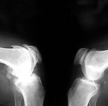
Visible osteophytes can be seen on radiographs, a narrowing of the joint space by 2-3 times compared to the norm, sclerosis of the subchondral bone and the formation of cystic cavities in the epiphyseal area.
Arthrosis of the 2nd degree is characterized by a decrease in the ability to work and the inability to perform certain types of work.
Arthrosis 3 degrees (the third stage of arthrosis)
arthrosis Stage 3 is a severe, advanced stage of the disease.At this stage, the following are observed:
- significant joint deformation (due to bone growth and fluid accumulation in the joint cavity);
- sharp limitation of movements, up to maintaining only rocking movements;
- sharp pains not only when moving, but also in a state of complete rest - constant pain accompanied by reflex spasms of nearby muscles, as well as the development of reactive synovitis;
- joint inflammation,
- common sensitivity to weather changes.
- the muscles around the knee are spasmed and atrophied;
The axis of the limbs is deformed, varus or valgus curvature of the legs is observed (that is, in the form of the letter "O" or "X").
In radiographs with arthrosis of the 3rd degree, an almost complete disappearance of the articular space, pronounced deformation of the articular surfaces and numerous marginal osteophytes are observed.Articular mice and calcification of pre-articular tissues can be detected.
In grade 3, the disease has progressed too far, and is often already the cause of permanent disability.It appears as follows:
- the pain becomes constant and painful: walking, and especially going up and down stairs, is a difficult trial for the patient;
- a loud cracking sound during every movement, clearly audible to others;
- joint deformation is severe, movements are limited only to a small amplitude or even impossible;
The photographs show the destruction of intra-articular structures (ligaments and menisci), as well as complete erosion of cartilage and signs of sclerosis (replacement of functional organs and structures with connective tissue).
Arthrosis 4 degrees
The state of complete destruction of the joint with arthrosisWhen the joint stops functioning completely, it is often classified as a special 4th degree of osteoarthritis.There is a so-called "joint block" - an acute pain syndrome in which even limited movement in the affected joint is impossible.The fourth degree of arthrosis is accompanied by unbearable pain in the joints, which cannot be relieved even with strong sedatives and intensive physiotherapy.Complete ankylosis (joint fusion) or neoarthrosis (formation of a false joint between the displaced ends of the bones) is possible.Independent movement in both cases is almost impossible.
Photographs show approximate sclerosis of the articulating surfaces with pronounced cystic elucidations, fusion of connecting bones in the area of the joint space.The development of the disease at this stage almost always means disability, which can only be prevented by the implantation of an artificial joint prosthesis.
Treatment of arthrosis
Treatment of arthrosis in the initial stage of the disease
It is better to start treatment of arthrosis as soon as possible, when the first signs appear - creaking in the joints, difficulty in movement.At this stage, drugs are useful - chondroprotectors that improve the structure of cartilage tissue, as well as vitamin and mineral complexes.
Physical therapy, proper nutrition and preventive measures are important.It should be noted that the prevention of arthrosis is of great importance to prevent the worsening of the disease.
Treatment of arthrosis 2 - 3 degrees
Although it is no longer possible to completely cure grade 2-3 arthrosis, the process of its development can be significantly slowed down.Treatment of arthrosis at this stage includes the following steps:
- relief or reduction of pain
- relief of inflammation in the joints.
- improve the restoration of cartilage tissue and slow down the degenerative processes in it.
In the acute period, treatment of arthrosis begins with pain relief.Non-hormonal anti-inflammatory drugs (NSAIDs) and analgesics are used for this purpose.Intra-articular injections of corticosteroids are possible.It is necessary to reduce the load on the joints;you should not walk or stand for a long time, or lift heavy objects.
After relieving the acute pain syndrome, the main task is to ensure, as much as possible, the activation of restoration processes in the joint itself and periarticular tissues: improving blood circulation, increasing metabolism, eliminating inflammatory processes.Chondroprotectors, vasodilators, as well as therapeutic exercises and physiotherapy are prescribed.
Treatment of grade 4 arthrosis
At this stage of the disease, the joint is almost completely destroyed.In this case, there is only one way out - surgery and replacement of the diseased joint with an endoprosthesis.The endoprosthesis significantly improves joint mobility and allows the patient to resume an active life, at least without pain.